
Objective
Intraoperative identification and mapping of cranial nerves remain some of the most challenging tasks in neurosurgery. Shortwave Infrared Orthogonal Polarization Imaging (SWIR-OPI) is an emerging technology that leverages significant water absorption at specific wavelengths to precisely detect and distinguish tissues with varying water content. Theoretically, nerves, which have lower water content, can be effectively distinguished from the higher water content environment of the cerebral cortex using SWIR-OPI. However, this technology has not yet been applied in any neurosurgical scenarios. This study investigates the potential application of SWIR-OPI in neurosurgery by examining its real-time imaging capabilities in fresh human and sheep cadaveric specimens.
Methods
SWIR-OPI images were collected from ten fresh, unembalmed human cadaveric specimens and five fresh sheep specimens. Anatomical landmarks, including cranial nerves, neural fibers, and cerebral cortex, were systematically evaluated. The Weber Contrast coefficient was employed to quantitatively analyze and compare the visibility of these structures within different environments.
Results
An exoscopy system equipped with SWIR-OPI, integrating high-performance SWIR light sources, linear polarizers, SWIR lenses, and InGaAs cameras, has been successfully established (Figure 1A). Analysis of SWIR-OPI images from cross-sectional sheep brains demonstrated that the 1550 nm wavelength exhibited the highest specificity for white matter tissues, with a significantly higher Weber Contrast coefficient compared to other short wavelengths (p<0.01, Figure 1B, C). Under 1550 nm SWIR-OPI, cranial nerves and fiber bundles (white matter) appeared as high-signal (white) regions due to their lower water content, while the cerebral cortex and nuclei (gray matter), with higher water content, appeared as low-signal (black) regions. The application of SWIR-OPI in cadaveric human heads revealed distinct and consistent patterns of high Weber Contrast coefficients in cranial nerves and fiber bundles compared to visible light imaging (p<0.01, Figure 1D, E). On the surface and cross-sections of the brainstem, SWIR-OPI images identified nuclei and gray matter contours that were indiscernible with standard visible light (Figure 1F-I).
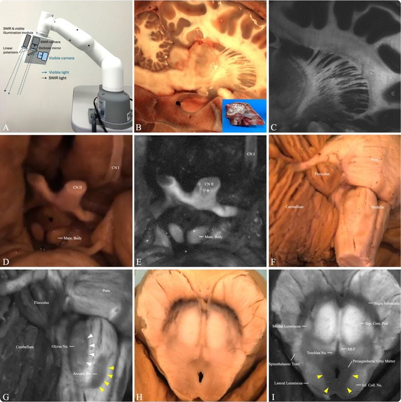
Exoscopy equipped with SWIR-OPI for nerve visualization on sheep and human specimen, revealing distinct and consistent high signals in cranial nerves and fiber bundles.
Conclusion:
SWIR-OPI provides clear, label-free, and real-time visualization of cranial nerves and fibers within surrounding cerebral structures, demonstrating significant potential for precise intraoperative nerve identification and mapping.
Authors
Stanford Hospital
Palo Alto, CA
Cision Vision
Mountain View, CA
Cision Vision
Mountain View, CA
Stanford Hospital
Palo Alto, CA
Stanford Hospital
Palo Alto, CA